Description
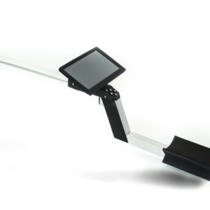
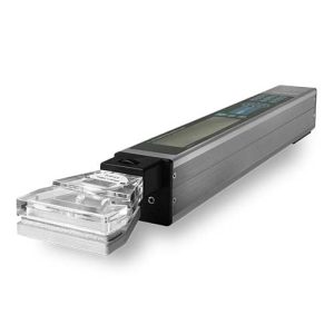
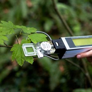
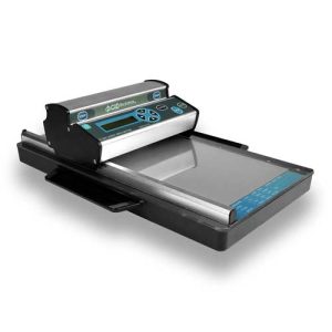
In-Situ Root Imager CI-600 from CID Bio-Science
High Resolution Root Scanning
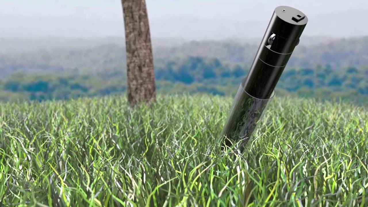
The study of fine root dynamics (production, turnover, and lifespan) and root system architecture (RSA) is at the forefront in the fields of ecology, agronomy, and plant breeding. Future gains in plant productivity will be driven by selection for traits that optimize acquisition of resources such as water and mineral nutrients under limiting conditions. The CI-600 leads the fine root-imaging field by offering researchers the ability to acquire high-resolution images of roots over time.
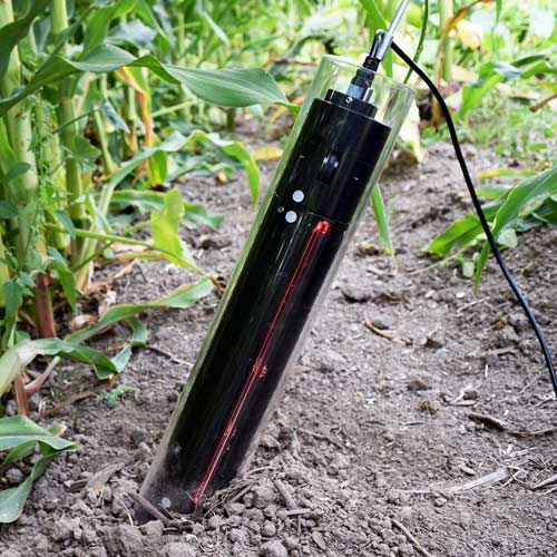
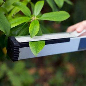
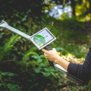
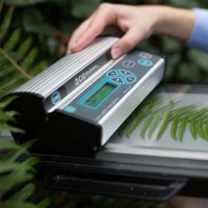
The CI-600 allows researchers the ability to scan multiple tubes in the field with one hand-held unit.
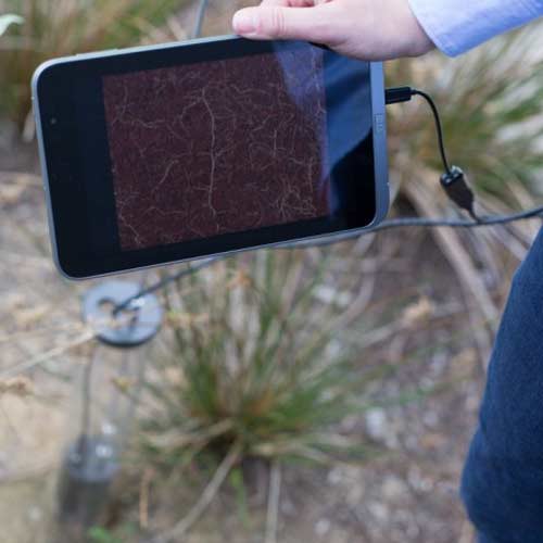
- Provides a high-resolution underground color image of living roots in the soil
- Enables observation of root system architecture and soil profile over time
Features
- High-resolution image – up to 23.5 million pixels
- Quick image capture – 0.5-4 minutes
- Linear scanning with no distortion
- Each scan generates a near 360 degres image (21.59 x 19.56cm) at up to 600dpi resolution
- Very portable and quick operation
- Enables observation of root system architecture and behavior during the entire growth season
- USB interface for laptop computer image storage
What’s in the Box
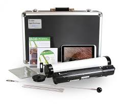
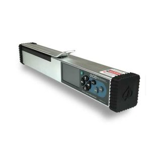
- Root imaging unit
- 3x 105cm clear tubes with end caps (custom length tubes available)
- Calibration tube
- Scanning software
- Operation manual
- Hardshell instrument case
- Tablet computer preloaded with RootSnap! image analysis software
Specifications
| Scanner Resolution: | 100, 300, & 600 DPI – up to 23.5 million pixels |
| Image Size: | 21.6 W x 19.6 L cm (8.5 W x 7.7 L in) |
| Scan Speed: | 0.5 – 4 minutes |
| Interface: | USB cable |
| Power Supply: | Tablet USB port |
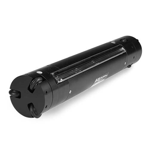
| Scan Head Dimensions: | 34.3 cm long x 6.4 cm diameter |
| Scanner Unit Weight: | 750 g |
Root Tube Dimensions
| Inner diameter: | 6.4 cm |
| Outer diameter: | 7 cm |
| Wall thickness: | 1.8 inch |
| Standard length: | 105 cm |
| Power Supply: | Tablet USB port |
CI-690 RootSnap! Root Image Analysis Software
Features
- Measures length, area, volume, & diameter
- Intuitive user interface
- Multi-touch screen functionality
- Integrated image enhancement
- Automated “Snap-to-Root” functionality
- Comprehensive data analysis package
- Time series analysis
- Files stored in common formats
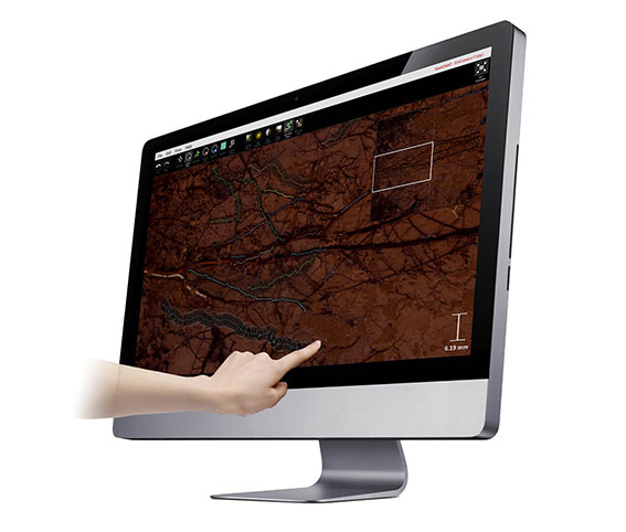
RootSnap! removes hours of tedious tracing by offering a revolutionary user interface that employs a multi-touch screen to easily trace roots using a finger. The software enables users to move quickly through root images to quantify root length, area, volume and diameter. The software has tracing enhancements like the “Snap-to-Root” function which automatically moves root tracing points to the center of the root. Additionally, RootSnap! has integrated image enhancement features which enables users to optimize each scanned image for more accurate processing.
Monitor root growth, dynamics, taxonomy, morphology, and behavior over time with RootSnap! Simplify root mapping using the multi-touch screen and our proprietary “Snap-to-Root” functionality. The superior RootSnap! user interface is intuitive and efficient. It uses familiar commands to manipulate images and files and stores information in common file formats (XML) and supports exporting data to Excel.
Benefits
- Easy to use
- Map roots quickly and efficiently
- Trace roots with a finger
- Easily manipulate images (colors, sharpness, contrast, etc.)
- Designed to work with Excel
- Monitor root growth, disease and behavior over time

 Français

Reviews
There are no reviews yet.